
Types of Microscopes
In the realm of scientific research, understanding the types of microscopes available is crucial for observing specimens at varying scales. Microscopes, integral tools in biology, chemistry, and medical research, enable scientists to investigate the structure and function of cells, tissues, and materials. With advancements in technology, the diversity of microscopes has expanded significantly, each designed to address specific research needs. This comprehensive guide will explore the various types of microscopes, their unique features, and their applications in scientific research.
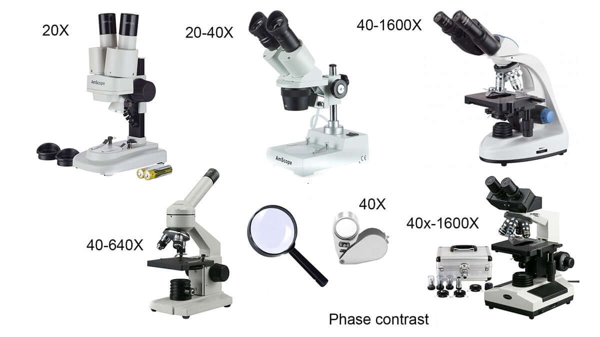
Types of microscope and their uses
Types of microscope and their uses

What are the Different Types of Microscopes?
There are different types of microscopes and each of these has different purposes of use. Some are suitable for biological applications, while others are used in educational institutions. There are also microscope types that find application in metallurgy and studying three-dimensional samples.
In this article, there are 5 such microscope types that are discussed along with their diagram, working principle and applications. These five Types of microscope and their uses are:
- Simple microscope
- Compound microscope
- Electron microscope
- Stereomicroscope
- Scanning probe microscope
Simple Microscope
A simple microscope is defined as the type of microscope that uses a single lens for the magnification of the sample. A simple microscope is a convex lens with a small focal length. The magnifying power of the simple microscope is given as:
|
|
Where,
- D is the least distinct vision
- F is the focal length of the convex lens
Simple Microscope Diagram

Principle of Simple Microscope
The working principle of a simple microscope is that when a sample is placed within the focus of the microscope, a virtual, erect and magnified image is obtained at the least distance of distinct vision from the eye that is held at the lens.
Application of Simple Microscope
- It is common among the watchmakers as they can view a magnified image of the smallest parts.
- It is also used by the jewellers for obtaining a magnified image of the fine parts of the jewellery.
- Most educational institutions such as schools and colleges use a simple microscope in their laboratories.
- Dermatologists (skin specialists) use simple microscopes to identify different skin diseases.
Compound Microscope
A compound microscope is defined as the type of microscope that has more than one lens. It has a combination of lenses and two optical parts known as an objective lens and eyepiece or ocular lens. The magnifying power of the compound microscope is given as:
|
|
Where,
- D is the least distance of distinct vision
- L is the length of the microscope tube
- fo is the focal length of the objective lens
- fe is the focal length of the eyepiece
Compound Microscope Diagram

Principle of Compound Microscope
The working principle of the compound microscope is that the combination of lenses enhances the magnification of the sample. The sample is first viewed as a primary image in the tube and viewed again in the eyepiece.
Applications of Compound Microscope
- The study of bacteria and viruses is possible with the help of a compound microscope.
- A compound microscope finds application in forensic laboratories.
- It is also used in metallurgy.
Electron Microscope
An electron microscope is defined as the type of microscope in which the source of illumination is the beam of accelerated electrons. It is a special type of microscope with a high resolution of images as the images can be magnified in nanometers.
There are two types of electron microscopes:
- The transmission electron microscope (TEM)
- The scanning electron microscope (SEM)

Principle of Electron Microscope
The metal used in an electron microscope is tungsten. A high voltage current is applied which results in the excitation of the electrons in the form of a continuous stream that is used as a beam of light. The lenses used in the electron microscope are magnetic coils. These magnetic coils are capable of focusing the electron beam on the sample such that the sample gets illuminated. As the flow of current increases, the strength of the magnetic lens increases. The electron beam flow is designed such that it cannot pass through the glass lens.
Application of Electron Microscope
- Quality control and failure analysis in industries are done with the help of an electron microscope.
- The images obtained in an electron microscope can be captured as electron micrographs with the help of specialized cameras.
- The study of metals and crystals became easy with the introduction of an electron microscope.
Stereo Microscope
A stereo microscope is defined as a type of microscope that provides a three-dimensional view of a specimen. It is also known as a dissecting microscope. In a stereo microscope, there are separate objective lenses and eyepiece such that there are two separate optical paths for each eye.
Stereo Microscope Diagram

Principle of Stereo Microscope
A stereo microscope works on the reflected light from the sample. The magnification of the microscope takes place at low power and hence, it is suitable for magnifying the opaque objects. It is suitable for thick and solid samples because it uses light reflected from the sample. The magnification of the stereo microscope is between 20x and 50x.
Applications of Stereo Microscope
- Examination of historic coins and artefacts is possible with the help of the stereomicroscope.
- It finds application in microsurgery.
- Viewing of crystals became easy with the use of a stereomicroscope.
Scanning Probe Microscope
The scanning probe microscope is defined as the type of microscope that finds applications in industries where the examination of the specimen is done at the nanoscale levels. The study of a specimen’s properties, its reaction time and its behaviour when stimulated can be done with the help of a scanning probe microscope.
Scanning Probe Microscope Diagram

Principle of Scanning Probe Microscope
The scanning probe microscope has a probe tip that is mounted on the end of a cantilever. The tip is so sharp that it can move precisely and accurately across the surface of the sample scanning every atom. The tip is placed close to the surface of the sample, such that the cantilever experiences a deflection due to forces. This deflection distance is measured by the laser. The final image after scanning is obtained on the computer.
Application of Scanning Probe Microscope
- It is used in studying different properties of the sample such as electrical properties.
- The magnetic property of the sample is studied using this microscope.
- The transferring of information on the sample can be done with the help of this microscope.
Related articles:
Stay tuned with BYJU’S to learn more about other concepts of Physics.
Frequently Asked Questions types of microscope and their uses – FAQs
What is the difference between low-powered and high-powered microscopes?
The basic difference between low-powered and high-powered microscopes is that a high power microscope is used for resolving smaller features as the objective lenses have great magnification. However, the depth of focus is greatest for low powered objectives. As the power is switched to higher, the depth of focus reduces.
What is depth of focus in a microscope?
The depth of focus in a microscope is defined as the distance between the objective lens and the sample plane. The depth of focus varies from person to person and is also dependent on the quality of focus.
What is the depth of field in a microscope?
The depth of field in a microscope is defined as the distance from the nearest object plane in focus to the farthest plane in the same focus. In microscopes, the depth of field is very short and is measured in units of microns.
What is the field of view in a microscope?
The field of view in a microscope is defined as the diameter of the illuminated circle which is seen through the eyepiece. With an increase in the magnification, there is a decrease in the field of view.
What is diopter adjustment?
The difference in vision between the two eyes is corrected with the help of diopter adjustment. Through diopter adjustment, the focus of individual eyepiece can be done so that the eyes feel comfortable while observing the sample.
Type of microscope image and description watch.
11 Different Types of Microscopes
The basic principle remains the same: microscopes use lenses to magnify objects, allowing researchers to see details that are invisible to the naked eye. However, the differences among types of microscopes lie in their design and the methods they employ to achieve magnification. Here, we categorize the different types of microscopes into a few major types: optical (light) microscopes, electron microscopes, and scanning probe microscopes.
Optical Microscopes
Optical microscopes are perhaps the most common types of microscopes used in laboratories today. They utilize visible light and a series of glass lenses to magnify images of small objects. These microscopes can be simple, like a single lens magnifying glass, or more complex systems with multiple lenses in a compound microscope configuration.
One prime example of an optical microscope is the compound microscope. It features two or more lenses—an objective lens and an eyepiece. Light passes through the specimen, and the objective lens creates an enlarged image that the eyepiece further magnifies. This type of microscope is widely used in schools and laboratories for biology experiments, where cellular structures need to be examined.
Another type of optical microscope is the stereo microscope, which provides a three-dimensional view of the specimen. Unlike the compound microscope, which is used for thin sections, the stereo microscope is ideal for dissecting larger samples, such as insects or flowers, providing a detailed view of surface structures.
Electron Microscopes
Electron microscopes represent a groundbreaking advancement in microscopy, allowing researchers to observe specimens at an atomic level. Unlike optical microscopes that use light, electron microscopes use a beam of electrons, which have much shorter wavelengths than visible light, providing vastly higher resolution images.
There are two primary types of electron microscopes: Transmission Electron Microscopes (TEM) and Scanning Electron Microscopes (SEM). TEM allows electrons to pass through the specimen, providing detailed internal structure images. It is commonly used in materials science and cellular biology, where understanding internal structures is crucial.
On the other hand, SEM provides detailed three-dimensional images of the surface of specimens. Samples must be coated with a conductive material if they are insulating. SEM is invaluable for examining large samples and providing topographical information about surfaces.
Scanning Probe Microscopes
Scanning Probe Microscopes (SPM) encompass a variety of techniques designed to measure surface properties at the nanoscale. Unlike optical and electron microscopes, SPM does not rely on light or electrons to generate images. Instead, it uses probes to scan the surface of a specimen, allowing researchers to visualize and manipulate materials at an atomic level.
The most well-known type of SPM is the Atomic Force Microscope (AFM), which uses a cantilever with a sharp tip to measure forces between the tip and the surface. AFM can provide high-resolution images and is valuable in studying biological samples, polymers, and nanostructures. It can also provide information on mechanical properties and surface textures.
Another type of SPM is the Scanning Tunneling Microscope (STM), which is particularly useful for studying conductive materials. It uses quantum tunneling to create images and can manipulate individual atoms on surfaces. STM has significantly contributed to nanotechnology and material science.
Specialized Microscopes
Beyond these primary categories, many types of microscopes have been developed for specific applications. For instance, fluorescence microscopes use specific wavelengths of light to excite fluorescent dyes attached to biological specimens, allowing researchers to study specific proteins and other molecules within cells.
Confocal microscopes offer improved image resolution and contrast by using a laser to illuminate the specimen, gathering data from a single point at a time, which reduces out-of-focus light. They are widely used in cell biology and histology for detailed imaging of samples.
Live-cell imaging microscopes enable the observation of live cells over extended periods, crucial for studying dynamic processes in real-time. Additionally, phase contrast and differential interference contrast (DIC) microscopes allow scientists to visualize transparent samples without extensive staining, preserving the natural state of live specimens.
Conclusion
Determining the right type of microscope for specific research purposes depends on various factors, including the nature of the specimen, the required resolution, and the specific details that need to be examined. Understanding the different types of microscopes and their functionalities is essential for researchers to select the appropriate tool for their investigations. With a plethora of available options, modern microscopists have the tools necessary to make significant discoveries across disciplines, pushing the boundaries of what science can reveal about the microscopic world.


